Actinic Keratosis: Recognition, Risk, and the Case for Individualized Treatment
A clinical reflection on AK definition and risk factors, the role of photodynamic therapy (PDT), PDT workflow considerations, and the use of cryosurgery for discrete lesions.
By Alyssa Ortega, PA-C, Alyssa Ortega, Physician Assistant | August 12, 2026
Actinic keratosis (AK) is one of the most common things we see in dermatology, but its clinical significance is routinely underestimated by patients and sometimes even by clinicians outside of dermatology. An individualized approach to AKs starts with accurate recognition, risk stratification, and thoughtful treatment selection. As a PA working in dermatology, the following is a clinical reflection on AK definition and risk factors, the role of photodynamic therapy (PDT), PDT workflow considerations, and the use of cryosurgery for discrete lesions.
Defining Actinic Keratosis and Contextualizing Risk
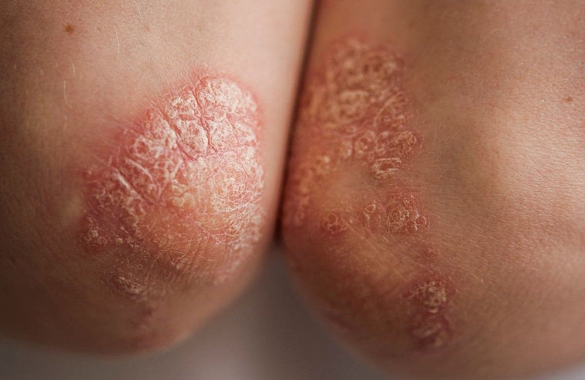
AKs present as erythematous, hyperkeratotic papules on chronically sun-exposed skin, commonly affecting the scalp, face, dorsal hands, and forearms. AKs are correctly positioned as precancerous lesions, and in my experience this framing is what actually gets patients to take treatment seriously. In my clinical practice, patients frequently dismiss these lesions as “dry skin” or “age spots,” which often delays evaluation and treatment.
It is important to emphasize the risk of malignant transformation at every patient encounter. A 10-year cohort study of over 17,000 patients found that individuals diagnosed with AK had more than 5 times the overall risk of developing skin cancer compared to matched controls, with the risk of squamous cell carcinoma (SCC), specifically being nearly eight times higher 1. Notably, there are no dermatoscopic features or histopathologic features that reliably predict which AKs will progress to SCC. Therefore, treatment of all lesions is prudent and considered the standard of care 2.
The risk of any single AK becoming an invasive SCC is something the literature has not fully settled. Rates vary depending on the study and the population, ranging from less than 0.1% per lesion per year to considerably higher in more aggressive disease. A secondary analysis of a randomized clinical trial put the 4-year cumulative cSCC risk at 3.7% overall, with that number jumping to 20.9% in patients with thick, hyperkeratotic lesions and 33.5% in those same patients who required additional treatment 3.
The presence of numerous AKs should prompt a conversation about field cancerization. It is also important to consider the concept that visible lesions exist within the broader zone of subclinical UV-damaged skin. Treating only individual lesions without addressing the entire field potentially leaves the patient at ongoing risk.
Photodynamic Therapy Selection
PDT’s mechanism of action is important to understand when selecting it as a treatment option. The photosensitizing agent, often aminolaevulinic acid (ALA), is selectively absorbed by abnormal keratinocytes. Upon activation by light, reactive oxygen species are generated, inducing targeted cell death while leaving surrounding healthy tissue largely intact.
From a clinical standpoint, PDT is advantageous for patients with multiple AKs in a confluent field. It offers excellent field treatment options with minimal scarring and strong patient satisfaction when setting appropriate expectations.
PDT Workflow
PDT workflow typically includes photosensitizer application, occlusion with plastic wrap or cling wrap, and incubation times ranging from 90 minutes to 3 hours.
A critical component of PDT success is postprocedural counseling. Patients need to avoid bright indoor lighting and UV exposure for 48 hours following treatment. Verbal and written instructions, along with sunscreen and sun-protective clothing recommendations, should be discussed at every PDT encounter.
Cryosurgery Selection
Cryosurgery remains a first-line treatment in patients presenting with isolated and discrete AKs rather than a broad field of photodamage. It is important to use freeze-thaw cycles of approximately 5 seconds per cycle and taking care to spare adjacent normal tissues. Multiple treatments may be necessary for thicker and resistant lesions. A clinical pearl to keep in mind is that any lesion that fails to respond to multiple cryotherapy sessions requires a biopsy to rule out SCC.
Conclusion
Understanding AK from recognition through treatment requires a clinically sound, practical framework. Our role extends beyond treating visible lesions; it includes educating patients on the significance of field cancerization, reinforcing photoprotection and maintaining a low threshold for biopsy when lesions respond atypically. Ultimately, choosing between PDT and cryosurgery, or both, comes down to knowing your patient, their lesion burden, their tolerance for downtime, and their history of adherence.
References
Guorgis G, Anderson CD, Lyth J, Falk M. Actinic keratosis diagnosis and increased risk of developing skin cancer: a 10-year cohort study of 17,651 patients in Sweden. Acta Derm Venereol. 2020;100(15):adv00128.
Criscione VD, Weinstock MA, Naylor MF, et al. Progression of actinic keratosis to squamous cell carcinoma revisited: clinical and treatment implications. Cancer. 2009;115(11):2523-2530.
Ahmady S, Jansen MHE, Nelemans PJ, et al. Risk of invasive cutaneous squamous cell carcinoma after different treatments for actinic keratosis: a secondary analysis of a randomized clinical trial. JAMA Dermatol. 2022;158(6):634-640.